By Dr. Grishma Kamble, MSK Senior Physiotherapist, Hayward
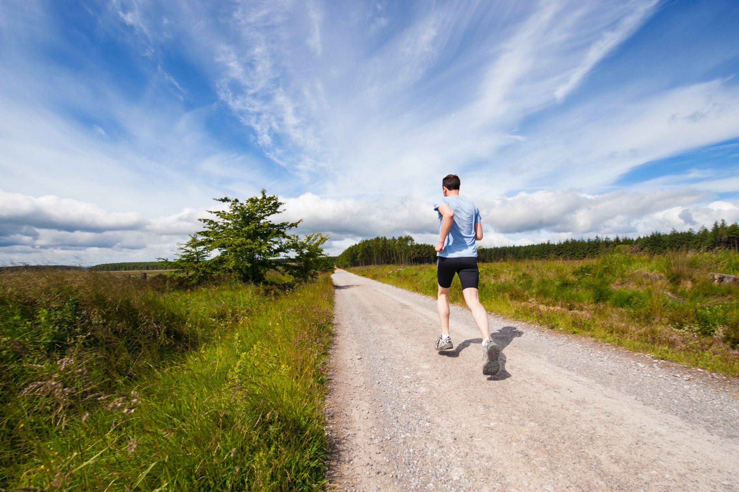
The management of lower limb injuries demands a precise understanding of biomechanics to address underlying dysfunctions effectively. In musculoskeletal (MSK) physiotherapy, biomechanics serves as the foundation for diagnosing movement impairments, designing targeted interventions, and achieving optimal patient outcomes.
Biomechanics and Lower Limb Pathology.
Biomechanics encompasses the analysis of forces acting on the body and their effects on movement. Lower limb injuries frequently arise from deviations in alignment, movement patterns, and load distribution. These dysfunctions, whether due to muscle imbalances, joint restrictions, or faulty kinematics, can lead to chronic pain, reduced performance, and recurrent injuries.
Key Pathologies Linked to Biomechanical Deficits
1. Patellofemoral Pain Syndrome (PFPS):
Aberrant patellar tracking, often secondary to weakness in the quadriceps and hip abductors, increases stress on the patellofemoral joint.
2. Achilles Tendinopathy:
Excessive load from poor ankle dorsiflexion mechanics or tight gastrocnemius-soleus muscles contributes to tendon degeneration.
3. Plantar Fasciitis:
Abnormal foot biomechanics, such as overpronation, alter load distribution on the plantar fascia, leading to microtrauma.
4. Iliotibial Band Syndrome (ITBS):
Caused by repetitive friction, ITBS is exacerbated by pelvic instability and poor lateral hip control.
Evidence-Based Biomechanical Interventions
1. Detailed Movement Analysis:
Gait and functional assessments are essential to identify abnormalities, such as asymmetrical weight-bearing, overpronation, or valgus collapse. These insights inform individualized treatment strategies.
2. Proximal Strengthening:
Weakness in proximal muscles, particularly the gluteus medius and maximus, often leads to compensatory loading. Focused strengthening improves joint stability and reduces abnormal stresses.
3. Manual Therapy Techniques:
Joint mobilization and soft tissue manipulation restore normal arthrokinematics, reduce pain, and improve movement efficiency.
4. Customized Orthotics and Footwear:
Tailored interventions address foot biomechanics to enhance load distribution and reduce strain on affected structures.
5. Neuromuscular Control Training:
Proprioceptive and motor control exercises, such as single-leg squats and balance drills, promote functional stability and injury prevention.
Case Study: Biomechanics in Practice
Patient Profile: A 35-year-old recreational runner presented with lateral knee pain persisting for 8 weeks.
Assessment Findings:
Gait analysis revealed excessive hip adduction and internal rotation during the stance phase, indicative of gluteus medius weakness. Secondary findings included tightness in the iliotibial band and reduced ankle dorsiflexion.
Intervention
Intervention:
Phase 1: Activation and strengthening of the gluteus medius using resistance band exercises.
Phase 2: Progression to functional, weight-bearing drills, including lateral step-downs and side planks.
Phase 3: Manual therapy to address IT band tightness and ankle mobility.
Outcome: The patient resumed pain-free running within six weeks, demonstrating improved hip control and normalized gait mechanics.
Professional Implications
As MSK physiotherapists, integrating biomechanical principles into clinical practice enables us to move beyond symptom management to address root causes of dysfunction. By leveraging advanced assessment tools and evidence-based interventions, we not only enhance recovery but also empower patients to achieve sustainable improvements in movement and performance.
Dr. Grishma Kamble